Eyeworld Weekly Update |
Volume 21, Number 12 |
25 March 2016 |
- Eye-tracking device for concussion detection granted U.S. approval
- First patients dosed in phase 1/2 RP study
- Foods high in vitamin C may cut cataract progression
- Mitochondrial dysfunction linked to Fuchs'
Eye-tracking device for concussion detection granted U.S. approval
The U.S. Food and Drug Administration granted regulatory approval for the Eye-Sync, an integrated head-mounted eye-tracking device that analyzes eye movement impairment through the use of virtual reality and may be able to determine if a player has suffered a concussion in less than 60 seconds, said developer SyncThink (Boston).
Rather than diagnose a concussion on the playing field, this device "detects disruption in visual information," the company noted in a press release. In January, the National Football League noted the rate of concussions in the 2015 season was up nearly 32% compared with data from 2014, while the Centers for Disease Control and Prevention reports that each year nearly 500,000 children are treated for a traumatic brain injury, including concussion.
First patients dosed in phase 1/2 RP study
A phase 1/2 study evaluating RST-001, a first-in-class gene therapy application of optogenetics for the treatment of retinitis pigmentosa (RP), has dosed its first patient, developer RetroSense Therapeutics (Ann Arbor, Michigan) said in a news release.
An initial dose-ranging study is proposed whereby 3 dose levels of RST-001 will be studied in 3 separate groups of adult patients with advanced disease. This first part of the study is aimed at determining a single dose of the experimental agent that is safe and well tolerated, to further evaluate in a fourth group of patients. The second part of the study is aimed at obtaining additional safety data at the highest tolerated dose and providing important additional clinical data to guide the design of future efficacy studies.
In 2014, the Food and Drug Administration granted orphan drug designation for RST-001 based on its development as a treatment of RP
Foods high in vitamin C may cut cataract progression
A diet rich in vitamin C could cut risk of cataract progression by a third, according to the American Academy of Ophthalmology (San Francisco), which published the study. The research is the first to show that diet and lifestyle may play a greater role than genetics in cataract development and severity.
Researchers at King's College London looked at whether certain nutrients from food or supplements could help prevent cataract progression, and examined more than 1,000 pairs of female twins from the U.K. Participants answered a food questionnaire to track the intake of vitamin C and other nutrients, including vitamins A, B, D, E, copper, manganese, and zinc. To measure the progression of cataracts, digital imaging was used to check the opacity of their lenses at around age 60. The researchers performed a follow-up measurement on 324 pairs of the twins about 10 years later.
During the baseline measurement, diets rich in vitamin C were associated with a 20% risk reduction for cataract. After 10 years, researchers found that women who reported consuming more vitamin C-rich foods had a 33% risk reduction of cataract progression. These results make the study the first to suggest that genetic factors may be less important in progression of cataract than previously thought.
Mitochondrial dysfunction linked to Fuchs'
Researchers at Schepens Eye Research Institute of Massachusetts Eye and Ear (Boston) have shown a link between mitochondrial dysfunction in corneal endothelial cells and the development of Fuchs' endothelial corneal dystrophy (FECD). This study is the first to demonstrate that lifelong accumulation of oxidative DNA damage leads to mitochondrial dysfunction and subsequent cell death in the tissue of the corneal endothelium. These changes are the result of free radical-induced molecular changes that are characteristic of FECD, the authors said.
In their study, the researchers detected an increase in mitochondrial and nuclear DNA damage in FECD and correlated the damage with loss of mitochondrial energy production. The result was mitochondrial fragmentation followed by release of cytochrome c, a small protein associated with the inner membrane of the mitochondrion. These age-induced molecular changes underscore the degenerative aspects of FECD pathogenesis. Although surgical therapies are effective in treating FECD, they all involve tissue transplantation. The research team plans to develop novel cytoprotective and anti-aging therapies that could provide alternative and safer treatment options for FECD patients, the university said.
This work was supported by an NEI/NIH RO1 Ey020581, and an Alcon Research Institute Young Investigator Grant.
RESEARCH BRIEFS
 Licensed Publications |
Licensed through ASCRS American Society of Cataract and Refractive Surgery, 4000 Legato Road, Suite 700, Fairfax, VA 22033-4003, USA.
All rights reserved. The ideas and opinions expressed in EyeWorld Asia-Pacific Weekly News do not necessarily reflect those of the ASCRS�ASOA or APACRS. Mention of products or services does not constitute an endorsement by the ASCRS�ASOA or APACRS. Copyright 2008, EyeWorld News Service, a division of ASCRS Media. |



 EyeSustain Update
 2024 APACRS TRAVEL GRANT
 Digital EyeWorld
 VOL. 39 (2023), ISSUE 3
  Membership Information
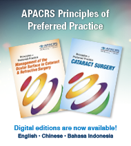 APACRS Principles of Preferred Practice